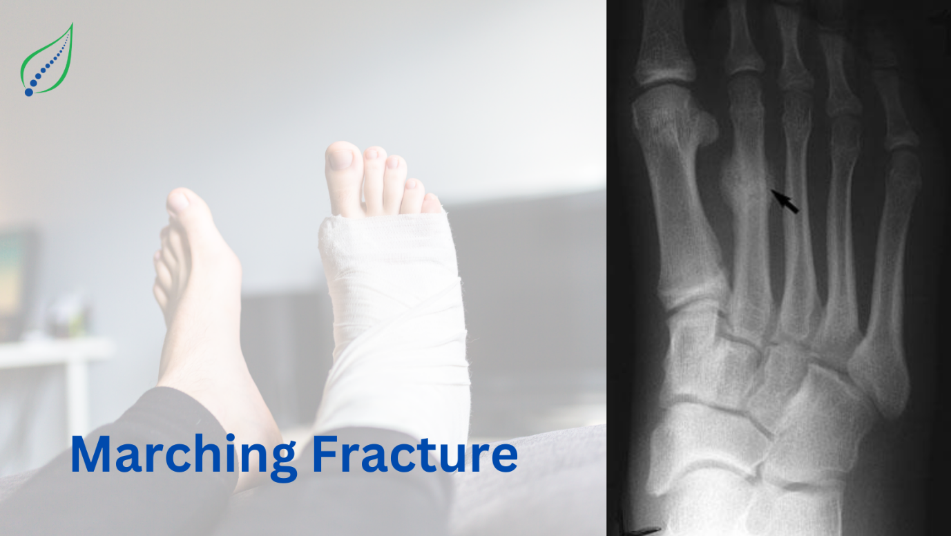
Marching Fracture
Repetitive stress placed over the metatarsal head leads to marching fracture. The term was coined in 1855 for foot pain and swelling experienced by Prussian soldiers while long marches.
25% incidence of these fractures commonly occur in athletes & military training. Condition is more commonly seen in females. People with past history of stress fractures are likely to develop.
CAUSES:
These metatarsal stress fractures, most commonly involve the second and third metatarsals. The repetitive impact on these metatarsal heads with weight-bearing exercises causes micro fractures, which consolidate into stress fractures and, therefore, are not a result of a single traumatic event.
Reason why the second metatarsal is most commonly involved:
- Second metatarsal neck is less flexible due to its strong ligamentous attachment to the 1st & 2nd cuneiforms & is prone to torsional forces applied.
- It is the longest of all metatarsals thus subjected to more force.
PATHOPHYSIOLOGY:
Osteoblastic activity lags behind osteoclastic activity during initial increases of exercise stress, leading to an increased incidence of stress fractures.
March fractures occur secondary to bone fatigue wherein the normal bone is unable to resist excessive mechanical demands or bone insufficiency (normal load on an abnormal bone).
Intrinsic factors such as nutritional deficiencies of Vitamin D & calcium deficiencies increase the risk of fractures. However extrinsic factors also play a major role, these include type of training & shoes.
Two types of stress fractures have been identified depending upon the location-
- Compressive
- Tensile.
Compressive stress fracture corresponds to fracture line on concave bone surface. Whereas, tensile stress fractures usually occur on the convex side of stressed bone.
SYMPTOMS:
- Pain improves with rest but increases again with activity.
- Quality of pain- Dull Aching
INVESTIGATIONS:
Physical examination should include- Observation of the painful site & of gait while weight bearing, Palpation of painful site eliciting any bony tenderness, examining joint movement & ranges.
Diagnostic imaging- Radiographs, MRI, Bone scans
In order to rule out intrinsic factors responsible for occurrence stress fractures, serum 25 (OH) vitamin D concentration can be considered in those suspected of nutritional deficiencies.
TREATMENT:
- Rest & icing are implemented for pain & swelling.
- Medicines- Analgesics.
- Orthopaedic boots can be used.
- Immobilization is often unnecessary. Weight bearing is allowed within limits of pain.
- Low impact exercises can be performed which involve- cycling, swimming, etc.
- Special considerations should be considered when fractures are present on the base of the fifth metatarsal and the neck of the second metatarsal. Management of such fractures include non-weight bearing with casts & crutches or surgical fixation in cases on non-union.
- Physical Therapy- Gait training & gentle stretches.
- Nutritional intake of Vitamin D & calcium
- Shock-absorbing insoles for shoes can distribute force during weight-bearing exercises.
- The type of training activity, surface, and intensity can be adjusted.
- When the patient is fully weight-bearing and pain-free with low-intensity exercise, the patient gradually increases their exercise intensity by 10% every week to avoid further stress fractures.


